Why not physical wall?
- Diameter of cells are different
- Wall may be too thin
- Choice of material
Scattering
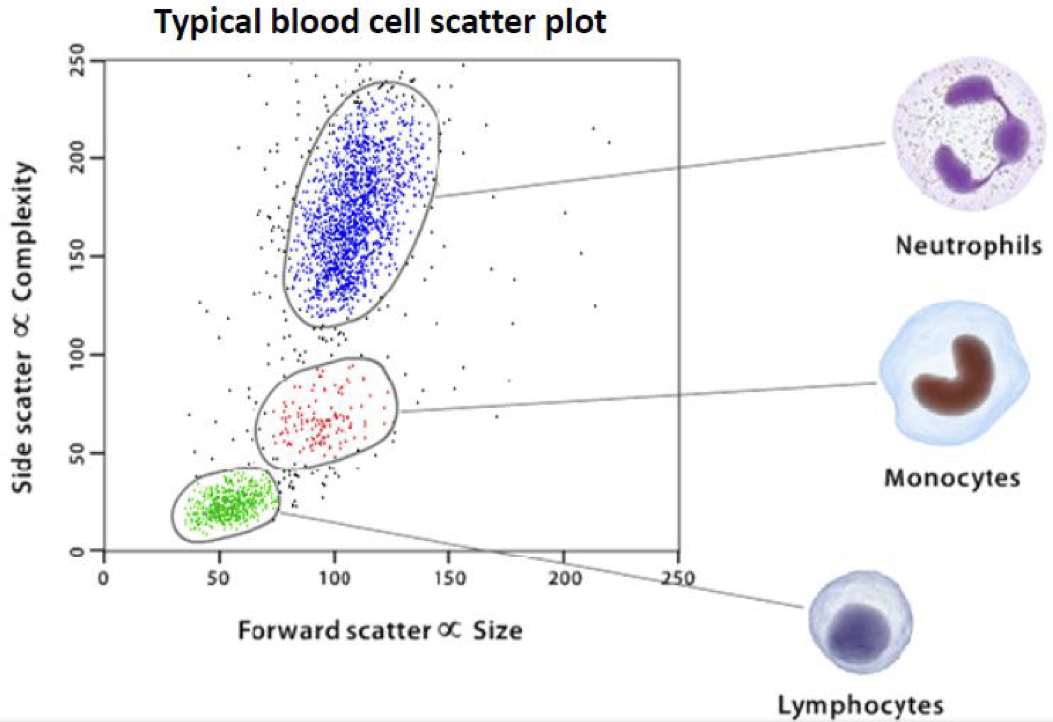
Forward Scatter (FSC)
- cell size
- Obscuration bar is used to block light when there is no cell
Side Scatter (SSC)
- granularity and complexity
- Detected at
2D Scatter Plot
Immunophenotyping
Flow cytometry can be used to distinguish between healthy and diseased cells. (e.g. myelomas, lymphomas, leukemias)
Fluorescence Imaging
Fluorochromes
Absorbs light energy excitation
Re-emit at a longer wavelength emission (in all direction)
The process of emission (fluorescence) is in the degree of
By conservation of energy, the emitted photons must have a lower energy.
In order words, lower frequency, longer wavelength).
e.g. blue light to green light
Excitation and Emission Spectra
Each fluorochrome has a unique excitation and emission spectra.
The emission spectra is always fixed.
Cellular Markers
- Voltage mathrmlse amount of fluorescence
Optical Filters
| Pass through | Dichroic |
|---|---|
| Absorb | Reflect |
| Short pass | Long pass | Band pass |
|---|---|---|
| e.g. | e.g. | e.g. |
Gating
- Direct the analysis software to consider only certain cell type.
- Produce additional histograms and dot plots to dissect subpomathrmlations, which have been labeled with fluorescent antibodies. (functional phenotyping)
Power of Cytometry
- Better understanding of immune mechanisms
- Correlation of cell analysis to genomics and proteomics
- fluorophores + FSC + SSC = parameters
- routine clinical use
- colors