What is imaging?
The science of extracting spatially and temporally resolved information at all physical scales.
Resolution
The smallest thing that can be seen.
The lower the number, the better the resolution.
Quality Characteristics
- Spatial resolution
- Details
- Contrast
- Noise
- Fidelity
Sensitivity and Specificity
| Actual True | Actual False | |
| Tested True | TP | FP |
| Tested False | FN | TN |
- High sensitivity sensitive in detecting abnormality good for abnormal patients
- High specificity not diagnosing normal as abnormal good for normal patients
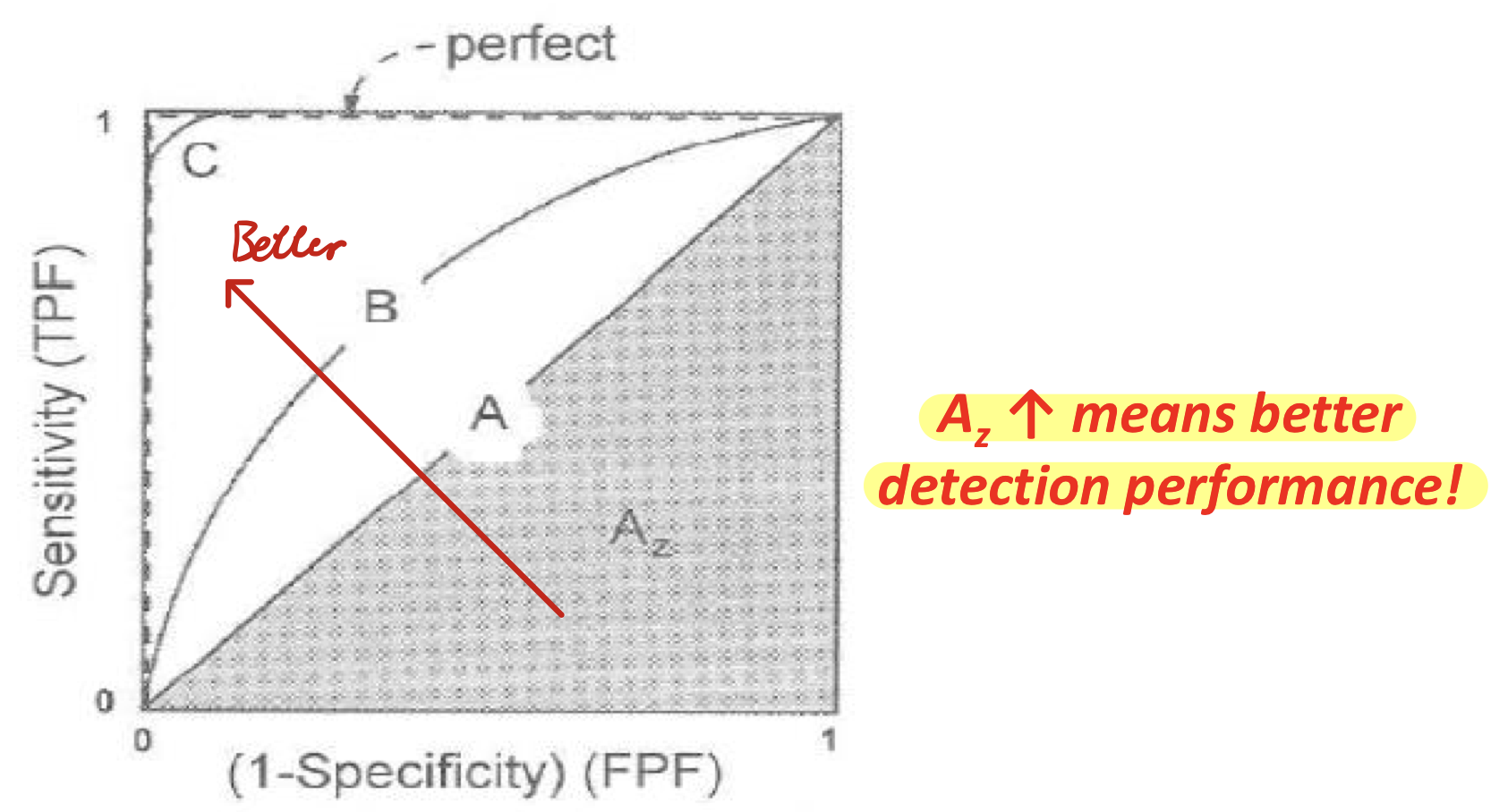
Receiver Operating Characteristics (ROC) Curve
X-ray
Production
- Current heats up cathode (filament)
- Cathode emit electrons
- Anode (tungsten) attracts high speed electrons
- The electron hits a tungsten atom
- An electron in a lower orbital falls out of the atom
- An electron in a higher orbital falls to the lower energy level
- Extra energy is released as X-ray
Linear Attenuation coefficient
Components
- Low-energy filter
- Collimator make X-ray parallel
- Collimating scatter grid
Applications
- Diagnostic imaging detection of structures and pathology of tissues (e.g. dental radiography)
- Orthopedics (bones)
- Chest
- Breasts
- Less penetrating, low energy source for soft tissues
- Fluoroscopy
- Moving images with X-ray
- Radiation therapy
Pros and Cons
Pros
- Obtain image of tissues inside body
- Detect diseases at early stage
- Widely accepts
- Low cost
- Quick
Cons
- A potential carcinogen (cancer)
- Soft tissues cannot be shown clearly
- Not recommended for pregnant women
- Image affected by position of patient and tissues
Computed Tomography (CT) Scan
Uses X-ray and computed tomography to obtain a 3D image of a body section.
Supplement X-ray and medical ultrasonography.
Gold standard in diagnosis.
Working Principle
- Measure 2D transmission projections at different angles, obtain 2D images around a single axis of rotation
- Obtain 3D image with digital geometry processing
| Single slide and axial scan | Helical (spirlal) scan |
|---|---|
| Scan one slice, translate table, repeat | Continuous linear movement between scans |
Pros and Cons
Pros
- Widely available
- Show bone changes best
- Detect life-threatening conditions
- Quick, painless, reasonably priced
- Only method that provides details images of bones, soft tissues, and blood vessels
Cons
- Radiation
- Misinterpret test results
- Allergic reaction to contrast material